Humerus Bone Anatomy
The humerus is the longest and largest bone of the upper limb․ It extends from the shoulder to the elbow, and plays a crucial role in arm movement․ This bone consists of a proximal end, a shaft, and a distal end, each with significant anatomical features․
Introduction
The humerus, the bone that forms the upper arm, is a vital component of the human skeletal system․ It connects the shoulder to the elbow, enabling a wide range of movements․ Understanding the anatomy of the humerus is crucial for healthcare professionals, especially those involved in orthopedic surgery, as it provides insights into potential injuries, treatments, and rehabilitation processes․ This comprehensive guide delves into the intricate details of the humerus, exploring its proximal and distal ends, shaft, and significant landmarks․
Proximal End
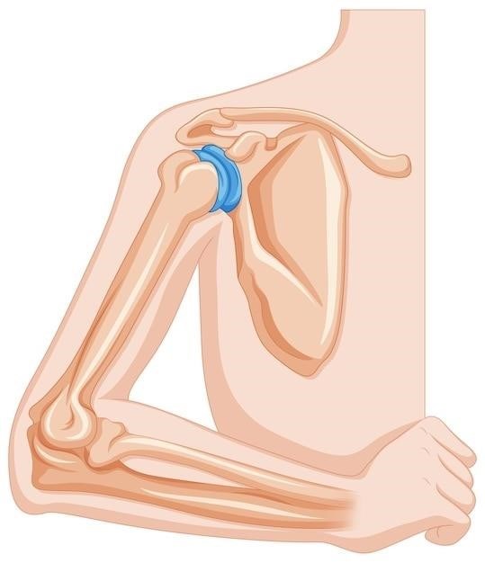
The proximal end of the humerus, the portion closest to the shoulder, is characterized by several prominent structures․ This region articulates with the scapula at the glenohumeral joint, forming the shoulder joint․ The head, a rounded projection, fits into the glenoid fossa of the scapula․ The anatomical neck, a constricted area below the head, serves as a point of reference for anatomical descriptions․ The surgical neck, located just below the anatomical neck, is a common site for fractures․
Head
The head of the humerus is a smooth, rounded projection that articulates with the glenoid fossa of the scapula, forming the glenohumeral joint, which is the shoulder joint․ This articulation allows for a wide range of motion in the shoulder, enabling activities such as reaching, throwing, and lifting․ The head is covered in hyaline cartilage, providing a smooth surface for joint movement․ It’s also slightly angled, contributing to the overall shape of the shoulder joint․
Anatomical Neck
The anatomical neck of the humerus is a constricted region located just below the head․ It serves as a boundary between the head and the greater and lesser tubercles․ The anatomical neck is a common site for fractures, particularly in older individuals due to the bone’s thinner structure in this area․ This region is also where several important muscles, such as the supraspinatus and infraspinatus, attach, contributing to shoulder stability and rotation․
Surgical Neck
The surgical neck of the humerus is a slightly constricted area located just distal to the greater and lesser tubercles․ It is named “surgical” because it is the most common site for humeral fractures, often resulting from falls or direct impact․ This location is particularly vulnerable due to its relatively thin structure and the forces that are exerted on it during arm movements․ The surgical neck is also the site of attachment for several important muscles, including the pectoralis major and latissimus dorsi․
Greater Tubercle
The greater tubercle is a prominent bony projection located on the lateral side of the proximal humerus․ It serves as an attachment point for several important muscles, including the supraspinatus, infraspinatus, and teres minor․ These muscles are responsible for external rotation and abduction of the arm․ The greater tubercle is easily palpable, and its position can be used as a landmark for locating other anatomical structures in the shoulder region․
Lesser Tubercle
The lesser tubercle is a smaller, more medially positioned bony projection on the proximal humerus․ It serves as the attachment point for the subscapularis muscle, which is responsible for internal rotation of the arm․ The lesser tubercle is less prominent than the greater tubercle and is often difficult to palpate․ However, it is an important landmark for understanding the anatomy of the shoulder joint and the muscles that control arm movement․
Intertubercular Sulcus
The intertubercular sulcus, also known as the bicipital groove, is a deep groove that separates the greater and lesser tubercles of the humerus․ It provides passage for the long head of the biceps brachii muscle tendon․ The intertubercular sulcus is lined by a synovial sheath, which helps to reduce friction as the tendon moves within the groove․ This groove is clinically significant because it can be a site of tendonitis, which can cause pain and restricted movement of the shoulder․

Shaft
The shaft of the humerus is the long, cylindrical portion of the bone that lies between the proximal and distal ends․ It is slightly curved, with a medial and a lateral side․ The shaft provides a broad surface for muscle attachment and is responsible for supporting the weight of the arm․ It also houses the nutrient foramen, a small opening that allows blood vessels to enter the bone and provide nourishment to the bone marrow․
Deltoid Tuberosity
The deltoid tuberosity is a roughened, triangular elevation located on the lateral surface of the humerus shaft․ This prominent feature serves as the attachment point for the powerful deltoid muscle, which is responsible for shoulder abduction (raising the arm away from the body)․ Its presence is significant for understanding the mechanics of shoulder movement and its potential role in fractures․
Radial Groove
The radial groove is a distinct, spiraling depression located on the posterior aspect of the humerus shaft․ This groove houses the radial nerve, a crucial nerve for controlling the muscles in the forearm and hand․ Its presence is important in understanding the potential for nerve injury in humerus fractures or dislocations, as damage to the radial nerve can result in weakness or paralysis in the hand and wrist․
Medial Epicondyle
The medial epicondyle is a prominent bony projection found on the medial side of the distal humerus․ It serves as the attachment point for several muscles that control wrist and finger flexion, including the flexor carpi radialis, flexor carpi ulnaris, palmaris longus, and pronator teres․ Due to its location and function, the medial epicondyle is susceptible to injury, particularly in athletes involved in repetitive overhead activities, leading to a condition known as medial epicondylitis or “golfer’s elbow․”
Lateral Epicondyle
The lateral epicondyle is a prominent bony projection located on the lateral side of the distal humerus․ It serves as the attachment point for several muscles that control wrist and finger extension, including the extensor carpi radialis brevis, extensor carpi ulnaris, extensor digitorum, and extensor digiti minimi․ Similar to the medial epicondyle, the lateral epicondyle can be prone to injury, particularly from repetitive wrist and finger extension movements, leading to a condition known as lateral epicondylitis or “tennis elbow․”
Distal End
The distal end of the humerus is characterized by its articulation with the radius and ulna bones of the forearm, forming the elbow joint․ This region features several important landmarks⁚ the capitulum, trochlea, coronoid fossa, olecranon fossa, and medial and lateral supracondylar ridges․ These structures play crucial roles in joint stability, movement, and muscle attachments․ The distal end of the humerus is also vulnerable to injuries, such as fractures and dislocations, which can significantly impact arm function․
Capitulum
The capitulum is a rounded, smooth articular surface located on the lateral aspect of the distal humerus․ This structure forms the articulation point for the head of the radius, contributing to the stability and mobility of the elbow joint․ The capitulum is essential for allowing the radius to rotate around the humerus, enabling movements like pronation and supination of the forearm․ It is also a crucial attachment point for several muscles, including the brachialis and biceps brachii, which play a vital role in elbow flexion․

Trochlea
The trochlea is a pulley-shaped articular surface located on the medial aspect of the distal humerus․ It articulates with the ulna, forming the hinge joint of the elbow․ This structure allows for flexion and extension of the forearm, crucial movements for everyday activities․ The trochlea’s shape helps guide the ulna during these movements, providing stability and limiting excessive motion․ It is also an attachment site for several muscles, including the triceps brachii, which extends the forearm․
Coronoid Fossa
The coronoid fossa is a shallow depression located on the anterior surface of the distal humerus, just above the trochlea․ This fossa serves as a crucial space for the coronoid process of the ulna to fit into during flexion of the elbow․ This process is a bony projection on the ulna that helps to stabilize the elbow joint during movement․ When the elbow is flexed, the coronoid process moves into the coronoid fossa, preventing the ulna from dislocating anteriorly․
Olecranon Fossa
The olecranon fossa is a deep depression located on the posterior surface of the distal humerus․ This fossa accommodates the olecranon process of the ulna during extension of the elbow․ The olecranon process is a large bony projection at the proximal end of the ulna, which serves as a lever arm for the triceps muscle․ When the elbow extends, the olecranon process moves into the olecranon fossa, providing stability to the elbow joint and preventing the ulna from dislocating posteriorly․
Medial and Lateral Supracondylar Ridges
The medial and lateral supracondylar ridges are prominent bony ridges located on the distal humerus, just above the medial and lateral epicondyles․ These ridges serve as attachment points for important muscles involved in elbow flexion and extension․ The medial supracondylar ridge provides attachment for the pronator teres muscle, which assists in pronation of the forearm․ The lateral supracondylar ridge provides attachment for the brachioradialis muscle, which assists in flexion of the forearm․
Clinical Significance
The humerus is a vital bone for arm function, making its anatomy clinically significant․ Fractures, dislocations, and other conditions can affect its structure and mobility․ Humeral fractures, especially those near the surgical neck, are common and can impact shoulder function․ Dislocations, often involving the glenohumeral joint, can cause pain and instability․ Conditions such as arthritis can also affect the humerus, leading to joint pain and stiffness․ Understanding the humerus’ anatomy is crucial for diagnosing and treating these conditions effectively․
Fractures
Humeral fractures are common injuries, particularly in the elderly and those involved in high-impact activities․ The surgical neck, a common fracture site, is particularly vulnerable due to its narrow diameter and proximity to the shoulder joint․ Other fracture locations include the shaft, the greater tubercle, and the distal end․ Treatment for humeral fractures varies depending on the severity and location of the break, ranging from non-surgical methods like immobilization to surgical interventions like open reduction and internal fixation․
Dislocations
Humeral dislocations, commonly known as shoulder dislocations, occur when the head of the humerus is displaced from the glenoid fossa of the scapula․ This injury is often caused by a direct blow to the shoulder or a forceful fall․ The most common type is an anterior dislocation, where the humeral head moves forward․ Symptoms include pain, swelling, and difficulty moving the arm․ Treatment typically involves reducing the dislocation, followed by immobilization to allow healing․
Other Conditions
Beyond fractures and dislocations, the humerus can be affected by a variety of other conditions․ These include⁚
- Epicondylitis⁚ Inflammation of the tendons around the medial or lateral epicondyles, causing pain and tenderness․
- Osteoarthritis⁚ Degeneration of the cartilage in the shoulder joint, leading to pain and stiffness․
- Bursitis⁚ Inflammation of the fluid-filled sacs (bursae) that cushion the shoulder joint, resulting in pain and swelling․
- Rotator cuff tears⁚ Damage to the tendons of the muscles that surround the shoulder joint, causing pain and weakness․
The humerus, as the longest bone in the upper limb, plays a pivotal role in arm function․ Understanding its anatomy, including the proximal, shaft, and distal regions, is crucial for comprehending the mechanics of arm movement and for diagnosing and treating various injuries and conditions affecting this bone․ From its articulation with the scapula at the shoulder joint to its connection with the radius and ulna at the elbow, the humerus serves as a fundamental structure for the upper extremity․


